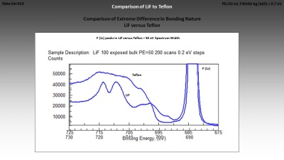
Brief Description
XPS Spectra Data Processing (SDP) Software
SDP v9.0 - 10 year License
$250
After installing please send the COMPUTER ID # shown by the installation screen,
+then we will return a 10 Year LICENSE NUMBER # that you must paste (add) to the installation screen.

DEMO Movies of SDP v8.0 (turn on sound)
Software Capabilities List
SDP v9 Installation Guide
Capabilities and Features
Advanced Peak-Fitting
Annotation (Add, Edit , Move)
ASCII Files Exported (Save As)
ASCII Files Imported
Atom % Quantification
Automated Charge Compensation
Backgrounds for Peak-Fitting
Binary Files Exported (Save As)
Binary Files Imported
Compatible Operating Systems
Depth Profile Display & Processing
Desk-Top-Publishing (DTP)
Display Functions
Drag & Drop to Open Files
Edit Sample Description / Names
Edit Spectrum Parameters
File Browsing
File Handling
Font Type & Style Control
Future Capabilities & Features
Graphic File Exporting
Keyboard Shortcuts
Line Thickness Control
Modify Raw Data
Multiple Spectra Display (MDI)
Multiple Spectra Printing
On-Screen Shortcuts
Overlaying of Multiple Spectra
Peak Finding (manual or automatic)
Peak Identification (manual or automatic)
Print Screen (2-10 screens on one page)
Printing and Page Layouts
RSF Values & BE Lookup Tables
Sample Description & System Name
Screen Capture (see Print Screen also)
Software Operation (Help menu)
Special Features
Spectral Lines Routine
Spectrum Parameters Menu
Capabilities included in Drop-Down Menus
Spectral Lines Menu
All 3 Spectral Line Columns are Scrollable
ASCII Tables for Spectral Lines can be Edited
BEs were derived from XI Library of XPS Spectra
“Chemical State Lookup Table” now available
Contains 1,100 Peaks (Peak Labels, RSF’s, Atom #)
Default Lookup Table is based on Aluminum X-rays
Default RSF values are based on Scofield calculations
Displays 20 lines of peak info in pop-up menu
Displays 3 columns: Peak Label, Element, & Atom #
Find & Identify Parameters adjusts the # of peaks found
Includes Charge Shift Correction (Offset) Slider
Peak Label shows BE, Element, Spin-Orbit, & RSF
Provides “Add Labels” and “Clear Lines”
Used for Auto Find & Identify Peaks from Conductors
Used to Manually Identify Peaks from Insulators
Useful to Identify Unexpected Peaks
Useful to Identify Weak Intensity Peaks
Advanced Peak-Fitting
Add Peaks (16) to Peak-Fit Table
Clear Peak-Fit Table
Define BE Shift Difference (8 pairs)
Define FWHMs
Define FWHM Ratios (8 pairs)
Define Gaussian:Lorentzian Peak-Shape Ratios
Define Peak Area Ratios (8 pairs)
Define Peak Asymmetry
Define Peak Energies
Define Peak Heights
Define Peak Height Ratio (8 pairs)
Define Peak Name (50 characters max.)
Define RSF value
Delete Any Peak from Peak-Fit
Display Peak-Fit Results On Top of Spectrum
Display Peak Error / Residual
Display Reduced Chi-Squared Result
Display Baseline Endpoint Values
Edit Peak-fit Results
Edit Peak Labels (50 character max.)
Fix (Lock) Binding Energies
Fix (Lock) FWHMs
Fix (Lock) Peak Heights
Fix (Lock) Peak Areas
Flat Background Subtraction / Addition
Link FWHMs (8 pairs)
Link Peak Areas (8 pairs)
Link Peak Energies (8 pairs)
Link Peak Heights (8 pairs)
Peak-fit Summary on Spectral Printout Page
Save Peak-Fit Results to Memory (use Save As)
Save Peak-Fit Table to Memory (use Save As)
Set Background Shape (Linear, Shirley, Tougaard)
Set BE Shift
Set Number of Peak-fit Iterations
Set Number of Points used to Calculate End-points
Set RSF exponent factor
Start Peak-Fitting
Stop Peak-Fitting
Zoom Display Range Graphically with Mouse
Zoom Display Range Numerically by X-Y Axes
Use Mouse Zoom to Define Peak FWHM & BE Max
Atom % Quantification
Adjust RSFs to correct for Instrument Effects
Adjust RSFs to correct for Kinetic Energy Effects
Atom % Summary on Spectral Printout Page
Change Exponent Factor to modify RSF values
Change Peaks used to Calculate Atom %
Clear Atom % Composition Table
Edit Atom % Composition Table
Measure New Peak Areas into Atom % Table
Modify RSF ASCII Lookup Table (As Needed)
Save Atom % Table to Permanent Memory
Use Other RSF Lookup Tables (Wagner etc.)
Desk-Top-Publishing (DTP)
Annotation (Add, Edit, or Move)
Atom % Summary on Spectral Printout Page
Capture “Tiled” Display by using “Alt-Print Screen”
Choose Color of Any Peak-Fit Line
Choose Color of Any Spectrum Line
Choose Font Style (Plain, Bold, Italic, Bold-Italic)
Choose Font Type of Axes and Peak Labels
Control Thickness of Spectrum and Peak-Fit Lines
Display / Hide Peak-fit or Sample Description Details
Display “Tiled” Multiple Spectra
Export Active Spectrum as Bitmap Image to Clipboard
Pass Energy and Charge Neutralizer on Spectrum
Paste Bitmap of Spectrum to Word, PowerPoint etc.
Peak-Fit Summary on Spectral Printout Page
Sample Description (2 lines) on Each Spectrum
Use “Paste Special” to Paste Captured “Tiled” Bitmap
ASCII Files Imported
CasaXPS (*.vms, *.txt)
Channel X-Y Generic (3 columns) (*.txt)
Grams 32 / Galactic (*.asc, *.txt)
JEOL 9000 (*.txt)
JEOL 7800 (*.vms, *.npl, *.txt)
JEOL 7810 (*.vms, *,npl, *.txt)
JEOL 7830 (*.vms, *.npl, *.txt)
Kratos Nova (*.des) saved as ASCII format
Kratos Ultima (*.des) saved as ASCII format
Kratos 165 (*.des) saved as ASCII format
Kratos HS (*.des) saved as ASCII format
Kratos Vision (*.des) saved as ASCII format
NPL VAMAS Standard DTF (*.vms)
NPL VAMAS Standard DTF (*.npl)
NRIM ComPro 3.0->6.1 (*.npl)
NRIM ISO-VAMAS Standard DTF (*.npl)
Omicron SPECTRA-Presenter (*.#)
Origin v6.0 and v7.0 (*.txt, *.asc)
PHI 5500, 5600, 5700, 5800 (*.asc with *.inf)
PHI Multi-Pak (*.asc)
PHI Quantum 2000 (*.asc)
PHI Quantera (*.asc)
RBD AugerScan v2 (*.txt)
Ron Unwin (*.#, *.txt)
Scienta (*.txt)
SPECS LHS (*.#)
SPECS Sage (*.exp) using 8.3 DOS names
SPECTRA-Presenter (*.#)
SSI DOS-Based M- or S-Probe (*.mrs, *.dpr, *.lpr, *.arp)
Synchrotron Light Sources (*.txt, *.asc)
VAMAS Standard DTF (*.vms)
VAMAS Standard DTF (*.txt)
VG ASCII (*.txt)
VG MicroTech VGX-900 (*.#)
VG version of VAMAS Standard DTF (*.txt)
Wild-Day (*.sp#)
XI-ASCII (*.mrs, *.dpr, *.lpr, *.arp))
X-Y Generic (2 columns) (*.txt)
OTHER Spectra able to be Imported
UPS
Sychrotron Light Sources
Linear Accelerator Light Sources
Fluorescence
UV-VIS
XRD
ASCII Files Exported (Save As)
Channel XY ASCII (*.asc, *.txt) for data analysis software
VAMAS Standard DTF (*.vms)
VAMAS Standard DTF (*.txt)
SSI-ASCII (*.mrs)
XI-ASCII (*.mrs)
XY ASCII (*.asc, *.txt) for data analysis software
Binary Files Imported
PHI MultiPak Binary Files (*.spe, *.pro)
SDP Binary Files (*.sdp)
SSI Binary Files (*.mrs)
Binary Files Exported (Save As)
SDP Binary Files (*.sdp, *.ovl)
SSI Binary Files (*.mrs, *.dpr, *.lpr)
Bitmap Graphic File Format is Exported for Desk-Top-Publishing (DTP)
The data in any “Active Display” (one spectrum or an overlay of several spectra) can be Exported as a Bitmap and then Pasted (Paste Special) or Imported by MS-Word, PageMaker, PowerPoint, Photoshop, etc..
Overlaying of Multiple Spectra
Auto-Scale Normalized Spectra
Copy Overlayed Spectra to New Window
Normalize Overlayed Spectra
Offset Overlayed Spectra
Proportionate Overlayed Spectra
Superimpose Overlayed Spectra
Zoom Overlayed Spectra
Backgrounds for Peak-Fitting
Auto-Subtract Linear Background
Choose Background (Linear, Shirley, or Tougaard)
Define # Points Used to Calculate BG Endpoints
Use Vertical Mouse Bar Cursors to Define Endpoints
Use Mouse Zoom to Define Endpoints
Drag & Drop to Open Files
Drag a compatible file onto an empty, open window and all spectra in that file are automatically imported and displayed
Drag a new datafile onto a filled windows and a new window is automatically generated.
File Handling
Change Data Directories
Change Disk Drives
Copy Files (use Windows commands)
Delete Active Spectrum
Delete Files (use Windows commands)
Drag & Drop Files to Open the File On-Screen
Export Spectra or Overlay as Bitmap
Merge Two or More Data-Files to an Open Data-File
Move File to New Directory (use Windows commands)
Open Any File Format Manually
Open Multi-Spectra Data-Files
Open Special File Formats Manually
Remembers Last Directory Opened
Save All Spectrum Modifications to Memory
Save Atom % Table to Memory
Select Spectrum from Multi-spectra File
File Browsing
By pressing the shortcut icons [ < > ] just under the main menu the user can display the next spectrum in a multi-spectra file.
By pressing the “Page Up” and “Page Down” keyboard keys the next spectrum in a multi-spectra data-file will appear.
Window Menu
“Tile” all open files to fit inside full window)
“Cascade” all open files
“Arrange Icons
Special Features
3D Montage Display of Depth Profile spectra
Adjust Exponent Factor that Modifies RSF Values
AES data processing
Annotation (add, edit and move)
Annotation (all annotations are saved to permanent memory!)
Automatic Find & Identify Peaks and Signals
Automated Charge Compensation
Auto Background Subtraction (Modify Counts)
Charge-up Correction (Offset) Slide
Chemical State Spectral Lines & Look-up Tables
Delete Individual Spectra from any File
Drag & Drop files to Open the File On-Screen
Icons as Short-cuts to make SDP easier-to-use
Import / Export VAMAS Standard ASCII DTF
Import / Export VAMAS Depth Profile spectra
Manually Find Peaks & Manually Identify Peaks
Merge Two or More Data-Files to an Open Data-File
Modify X and Y axis Labels (e.g. cts -> cps)
Modify Sample Description (2 lines) or System Name
Modify Spectrum Parameters (BEs, PE, Dwell, Scans
Reverse axis of KE or BE during import (Import Options)
Save All Atom % Tables to Permanent Memory
Save All Processed Spectra to Permanent Memory
Scaling and Zooming of Depth Profile 3D Montage
Peak Finding & Peak Identification
“Spectral Lines” lookup table is used to identify the peaks in any insulators or conductors.
Automated “Find & Identify” (Find & ID) for conductors
“Find Peaks” uses the pure element BE Lookup Table derived from the XI Spectral Library of Pure Elements
“Find Peaks” is useful for insulating materials due to charge up effects
“Adjust Find Peaks Parameters” adjusts the # of peaks found
Manual “Find Peaks” is used to find and integrate peaks of insulators
BE Lookup Table can be Used to locate and identify simple chemical state differences
“Spectral Lines” table is used to identify the peaks in any spectrum. Most Auger and Energy Loss signals are included.
The Spectral Lines tables can be modified by the user at any time.
Printing & Page Layouts
Adjust Printer Page Margins
Print “Landscape” or “Portrait” style
Print Active Display (Spectrum)
Print Atom % Composition Table (detailed)
Print Atom % Composition Table (simple)
Print Screen prints all spectra that are displayed
Print Spectra with Peak-Fits Data on same page
Print Wide Scans with Atom % Summary same page
Print Table of Peak-fit Parameters
Print to any Windows Compatible Printer
Print to different paper sizes (Letter, A4, B5, …)
Print Multiple Spectra in MS-Word by using the Tile
Select Windows Printer (Laser or Color Inkjet)
Window function together with the “Alt-Print Screen”
and “Paste” Bitmap function of Win
Compatible Operating Systems
Microsoft Windows 10, 8, 7, XP,
(supports long filenames)
Macintosh with Intel chip and Windows OS
(Not compatible with MS-DOS)
On-Screen Shortcuts
Add New Peak to Peak-Fit
Auto Find & Identify Peaks
Change to Next / Previous Spectrum
Clear XPS Spectral Lines from Screen
Define Background Shape
Display Peak Table
Display Peak-Fit Information Table
Display Peak-Fit Parameters
Display XPS Spectral Lines On-Screen
Start Peak-Fitting
Keyboard Shortcuts (Ctrl+ ?)
Ctrl+A “Adjust” data by some value (+ – / *)
Ctrl+C “Copy Bitmap” of active screen to Clipboard
Ctrl+D Display “Differentiation” menu
Ctrl+E Switch “Energy” scale (KE / BE)
Ctrl+F Auto “Find & Identify” peaks
Ctrl+G Display “Smoothing” menu
Ctrl+M “Merge” two data-files
Ctrl+N Edit “Names & Sample Description”
Ctrl+O Display “Open File” menu
Ctrl+P Display “Print” menu
Ctrl+S Display “Save As (VAMAS, ASCII, Binary): ” menu
Ctrl+T Display default “Tougaard” Background
Ctrl+X Display “XPS Spectral Lines” menu
Ctrl+Z “Un-Do last action
Alt+T Hide default “Tougaard” Background
Alt+X Hides XPS Spectral Lines already Added
Long File Names Supported
Open & Save Files with Long File Names
Charge-up Correction of Insulators
Adjust (+/-) XPS Spectral Lines for Charge-up
Semi-Automated Multi-Spectra Charge Correction
Shift BE by Editing BEs in Spectrum Parameters Menu
Modify Raw Data
Additive Smoothing or Differentiation
Revert to Original Data
Savitzky-Golay Differentiation (1-N pts)
Savitzky-Golay Smoothing (1-N pts)
Scale Raw Data by Number Factor
Start / Finish BE / KE values
Relative Sensitivity Factors & BE Tables
Add a Special RSF Table for UPS or SOR or …
Adjust RSFs to correct for Instrument Effects
Adjust RSFs to correct for Kinetic Energy Effects
Change Exponent Factor to modify RSF values
Change Scofield (Lookup) tables to any other Lookup table
Use any XPS RSF Tables (Al, Mg, Wagner, PHI, VG..)
Use any AES RSF Tables (10 KV, PHI, VG…)
(Note: RSF tables are combined with BE Lookup tables)
Display Functions
Auto-Scale Y-Axis automatically sets “Y” min-max
Chi-Squared Calculation is Displayed on Menu Bar
Choose Color of Any Peak-Fit Line
Choose Color of Any Spectrum Line
Choose Font Style (Plain, Bold, Italic, Bold-Italic)
Choose Font Type of Axes and Peak Labels
Choose Thickness of Spectrum and Peak-Fit Lines
Current Spectrum Number displayed on menu bar
Delete Peak Label and Peak Area from Wide Scan
Display/modify 2 lines of Sample Description
Display / Hide any part of Peak-fit or Atom% Tables
Display Date and Time of Data Collection
Display Import Format
Display Atom % Summary on Wide Scan
Display File Type or Operator Name
Display Pass Energy and Charge Neutralizer settings
Display Peak-fit Summary on Peak-fitted spectra
Display XPS Spectral Lines (Peaks) for all Elements
Display windows can be re-sized (Windows feature)
Double click on Menu Bar for full Screen View
Edit Two Lines of Sample Description
File-Path is displayed in Top Menu Bar
Manual Scaling of X-Y Axes
Modify X and Y axis Labels (e.g. cts -> cps)
Reset X-Y Axes
Reverse X axis by changing “Import Options”
Revert to original display
Second Description line shows file-path (if blank)
Step-by-Step “Un-Do”
Switch between KE and BE on X-axis
Tile, Cascade or Close All Open Files
Total Number of Spectra in File displayed in Menu Bar
X-axis displays as either KE or BE (eV) Scale
Y-axis displays as total Electron Counts Scale
Zoom (expand) Display by using Zoom Box
Zoom (expand) Display by using vertical cursors
Zoom / Un-Zoom X-Y Axes of Spectrum Displayed
Edit Spectrum Parameters Menu
Allows user to modify “Left X (BE/KE) value”
Allows user to modify “Right X (BE/KE) value”
Allows user to modify “Source Energy”
Allows user to modify “Pass Energy”
Allows user to modify “Dwell Time” (seconds)
Allows user to modify “Number of Scans”
Displays total number of data points in each spectrum
Displays “Left value” KE/BE of left most data point
Displays “Right value” KE/BE of right most data point
Displays Current “X” scale setting (KE or BE)
Displays “X-ray Source Energy” from original file
Displays “Pass Energy” reported in original file
Displays “Dwell Time” in seconds
Displays Number of “Scans” reported in original file
Displays default value = 1 if value is not available
Edit Sample Description / Names Menu
Displays System Name or Operator Name
Displays Two Lines of Sample Description
User can Edit System Name or Operator Name
User can Edit Both Lines of Sample Description